Plik:Gobiraptor.png
Rozmiar podglądu – 800 x 551 pikseli. Inne rozdzielczości: 320 x 220 pikseli | 640 x 441 pikseli | 1024 x 705 pikseli | 1280 x 881 pikseli | 1954 x 1345 pikseli.
Rozmiar pierwotny (1954 × 1345 pikseli, rozmiar pliku: 1,45 MB, typ MIME: image/png)
Opis
| Opis |
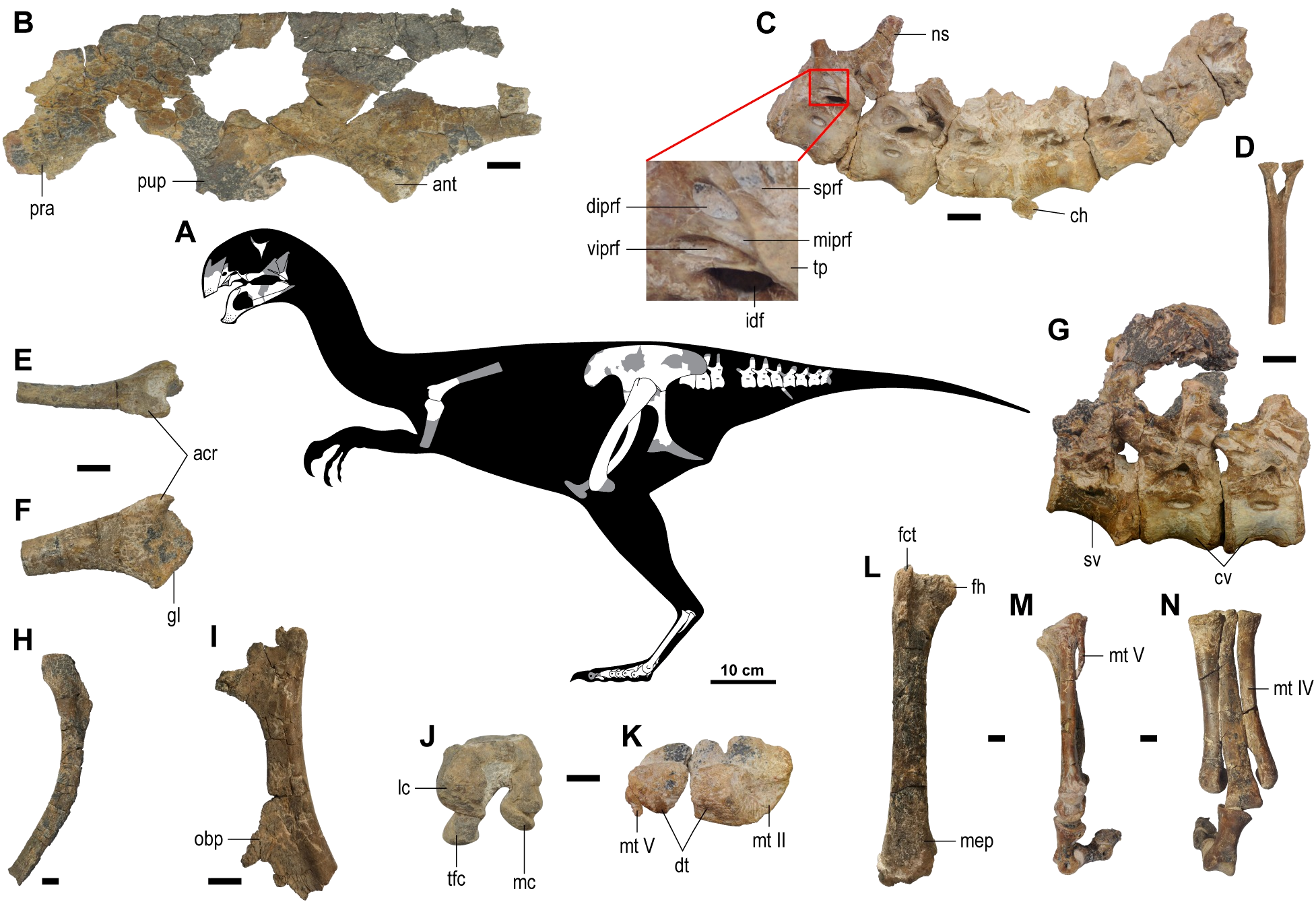
English: (A) Skeletal reconstruction in left lateral view (missing and damaged portions of the bones in gray). (B) Left ilium in lateral view. (C) Proximal caudal vertebrae in left lateral view with close-up of the infraprezygapophyses. (D) Chevron in cranial view. (E-F) Right scapula in dorsal (E) and lateral (F) views. (G) Last sacral and the two proximalmost caudals in left lateral view. (H) Right pubis in medial view. (I) Right ischium in lateral view. (J) Right femur in distal view. (K) Left metatarsus and distal tarsals in proximal view. (L) Right femur in cranial view. (M-N) Left metatarsus in lateral (M) and dorsal (N) views. Abbreviations: acr, acromion process; ant, antitrochanter; ch, chevron; cv, caudal vertebra(e); diprf, dorsal infraprezygapophyseal fossa; dt, distal tarsal(s); fct, cranial trochanter of femur; fh, femoral head; gl, glenoid fossa; idf, infradiapophyseal fossa; lc, lateral condyle; mc, medial condyle; mep, medial epicondyle; miprf, middle infraprezygapophyseal fossa; mt II, metatarsal II; mt IV, metatarsal IV; mt V, metatarsal V; ns, neural spine; obp, obturator process; pra, preacetabular process; pup, pubic peduncle; sprf, supraprezygapophyseal fossa; sv, sacral vertebra; tfc, tibiofibular crest; tp, transverse process; viprf, ventral infraprezygapophyseal fossa. Scale bars equal 10 cm in (A); 1 cm in (B-N).
https://doi.org/10.1371/journal.pone.0210867.g004 |
| Data | |
| Źródło | https://doi.org/10.1371/journal.pone.0210867.g004 |
| Autor | 2019 Lee et al. |
Licencja
Ten plik udostępniony jest na licencji Creative Commons Uznanie autorstwa 4.0 Międzynarodowe.
- Wolno:
- dzielić się – kopiować, rozpowszechniać, odtwarzać i wykonywać utwór
- modyfikować – tworzyć utwory zależne
- Na następujących warunkach:
- uznanie autorstwa – musisz określić autorstwo utworu, podać link do licencji, a także wskazać czy utwór został zmieniony. Możesz to zrobić w każdy rozsądny sposób, o ile nie będzie to sugerować, że licencjodawca popiera Ciebie lub Twoje użycie utworu.
Historia pliku
Kliknij na datę/czas, aby zobaczyć, jak plik wyglądał w tym czasie.
| Data i czas | Miniatura | Wymiary | Użytkownik | Opis | |
|---|---|---|---|---|---|
| aktualny | 02:12, 14 mar 2019 | | 1954 × 1345 (1,45 MB) | Ras67 | -background, opt. |
Wykorzystanie pliku
Poniższa strona odwołuje się do tego pliku: